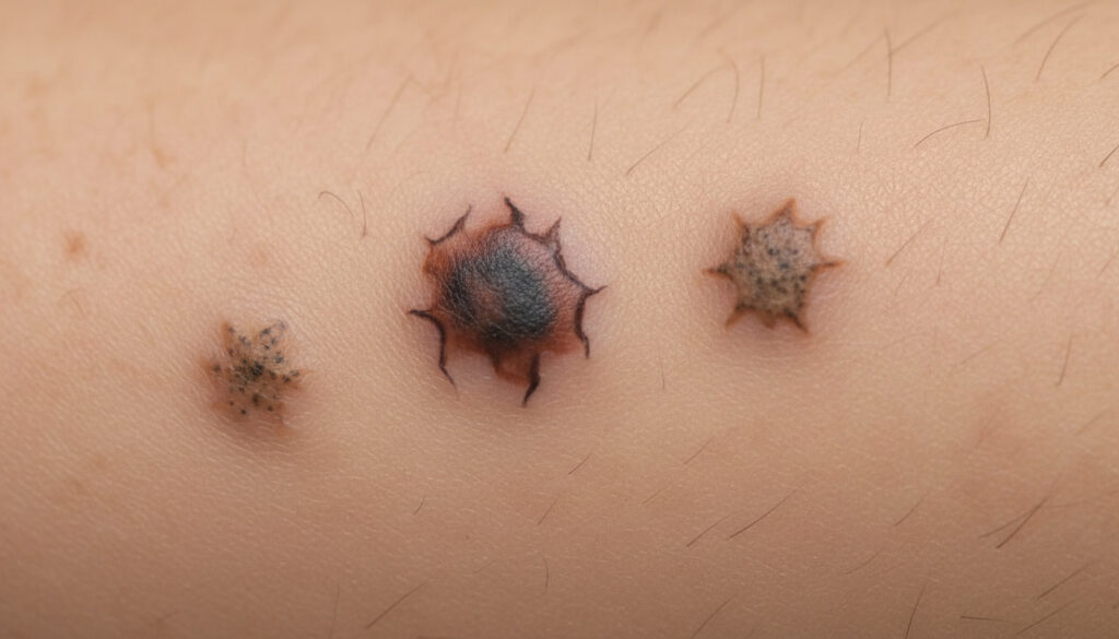
What Does Skin Cancer Look Like – Key Signs to Spot Early
The Visible Threat: Recognising Skin Cancer in Its Many Forms
Few discoveries provoke the immediate anxiety of finding an unfamiliar mark on your skin. While most spots remain harmless, understanding what malignancy actually looks like can mean the difference between a simple excision and aggressive treatment. Skin cancer rarely announces itself with obvious pain; instead, it communicates through subtle alterations in colour, texture, and symmetry that evolve over weeks or months.
The Visual Landscape: Three Distinct Presentations
Skin cancer manifests through three primary visual languages. Melanoma often appears as the “ugly duckling”—a mole that looks different from your other spots, potentially displaying variegated colours ranging from tan to black, sometimes with blue or white undertones. These lesions typically respect no uniform border, creating irregular, notched edges that blur into surrounding skin.
Basal cell carcinoma presents differently, frequently materialising as a waxy or pearly bump that might resemble a flesh-coloured mole or a persistent pink patch. You may notice visible blood vessels spiderwebbing across the surface, or the lesion may repeatedly crust and bleed without fully healing.
Squamous cell carcinoma tends toward the aggressive-looking: firm red nodules, flat lesions with scaly crusts, or rough patches that feel like sandpaper against your fingertips. Unlike benign dry skin, these areas persist and thicken despite moisturising.
Critical Warning Patterns
Dermatologists rely on systematic observation frameworks to distinguish benign from malignant. The ABCDE criteria remain the gold standard for melanoma detection: Asymmetry (one half unlike the other), Border irregularity, Colour variation, Diameter larger than six millimetres, and Evolution or change over time.
Beyond alphabetic mnemonics, the “ugly duckling” concept proves equally vital. In a field of uniform moles, one that stands out—darker, larger, or differently shaped—warrants professional evaluation regardless of other characteristics. For non-melanoma types, the American Academy of Dermatology emphasises persistent growth, spontaneous bleeding without trauma, and lesions that fail to heal within three weeks as primary red flags.
Comparative Identification Guide
| Type | Typical Appearance | Texture | Common Locations |
|---|---|---|---|
| Melanoma | Dark, multi-coloured patch; irregular borders | Smooth or slightly raised; may ulcerate | Back (men), legs (women), sun-exposed areas |
| Basal Cell Carcinoma | Pearly or waxy bump; pinkish patch | Firm, translucent; visible blood vessels | Face, ears, neck, scalp |
| Squamous Cell Carcinoma | Red nodule; scaly crust; open sore | Rough, sandpaper-like; may crust | Face, ears, hands, arms |
| Actinic Keratosis | Rough, scaly patch; flesh-toned or pink | Dry, gritty sensation | Face, scalp, backs of hands |
Detailed Clinical Characteristics
Melanoma demands particular vigilance regarding evolution. A spot that changes height, develops new colours, or begins itching and bleeding requires immediate assessment. Cancer Research UK notes that nodular melanoma specifically appears as a firm, dome-shaped growth that may be uniform in colour but grows rapidly over weeks.
Basal cell carcinomas, while rarely metastasising, display distinctive behavioural patterns. The superficial type appears as a reddish, slightly scaly patch that might resemble eczema, while morphoeic variants present as waxy, scar-like areas with indistinct borders that slowly expand.
Squamous cell carcinomas often emerge from precursor lesions called actinic keratoses—rough, dry patches caused by cumulative sun exposure. When these transform, they may develop into raised, horn-like growths or persistent ulcers that resist healing, particularly on the lip or ear where they carry higher metastatic potential.
Progression and Temporal Changes
Skin cancer rarely appears overnight. Melanomas typically evolve over months, progressing from flat, irregular macules to raised nodules as they penetrate deeper skin layers. Basal cell carcinomas grow slowly, often expanding imperceptibly over years while maintaining their characteristic pearly appearance.
Squamous cell lesions accelerate more quickly, potentially doubling in size over months. The NHS cautions that any sore failing to heal within four weeks, particularly one that repeatedly scabs and bleeds, requires urgent dermatological review regardless of initial appearance.
Diagnostic Clarity
Visual examination alone cannot confirm malignancy. Dermatologists employ dermoscopy—magnified light examination revealing subsurface structures invisible to the naked eye—to evaluate suspicious lesions. Asymmetric pigmentation patterns, blue-white veils, and irregular vascular distributions under magnification prompt biopsy.
However, certain mimics complicate identification. Seborrhoeic keratoses present as waxy, “stuck-on” brown growths that resemble melanoma but feature uniform surface textures. Pyogenic granulomas appear as rapidly growing red nodules that bleed easily, mimicking amelanotic melanoma. Professional evaluation remains essential when uncertainty persists.
Statistical Context and Detection Impact
The visual characteristics carry life-or-death significance. Mayo Clinic data indicates that five-year survival rates for melanoma exceed 99% when detected early while confined to the epidermis, dropping to 27% once distant metastasis occurs. Despite this, many individuals delay presentation until lesions become physically symptomatic.
Non-melanoma skin cancers, while less lethal, cause significant morbidity through local tissue destruction. Basal cell carcinomas specifically can infiltrate surrounding bone and cartilage if left untreated, despite their benign reputation.
Expert Perspectives
“The most dangerous misconception is that skin cancer always looks like a changing mole. We frequently see patients who ignored a persistent scaly patch or a pearly bump because it didn’t match the melanoma photographs they’d seen. Each skin cancer type speaks its own visual language.” For more information on skin cancer signs, check out Останні результати матчів.
— Dr. Sarah Chen, Consultant Dermatologist
“Patients should examine their skin monthly, looking not just for the ‘classic’ dark mole, but for anything that violates the pattern of their normal skin. Evolution—change over time—remains the single most important feature across all skin cancer types.”
— American Cancer Society Clinical Guidelines
Key Recognition Principles
Effective skin surveillance requires abandoning the search for a single “cancerous look.” Instead, practise pattern recognition and change detection. Document existing moles through photographs to establish baselines against which future changes can be measured. Pay particular attention to lesions in hard-to-see areas—scalp, back, soles of feet—where malignancies often progress unnoticed.
Remember that skin cancer affects all skin tones. While melanoma incidence rises with lighter pigmentation, acral lentiginous melanoma (appearing on palms, soles, and under nails) occurs disproportionately in darker skin and often presents as a longitudinal brown-to-black streak in the nail or an irregular dark patch on the sole.
Frequently Asked Questions
How can I tell the difference between a pimple and skin cancer?
Pimples typically resolve within one to two weeks, developing a white head before healing. Skin cancer persists beyond three to four weeks, may bleed spontaneously without trauma, and lacks the cyclical inflammation of acne. Basal cell carcinomas can resemble persistent pimples but feel waxy or pearly rather than tender and inflamed.
Does skin cancer always appear as a dark mole?
No. While melanomas often display dark pigmentation, amelanotic melanomas appear pink, red, or skin-coloured. Basal cell carcinomas frequently present as pearly or pink bumps, and squamous cell carcinomas may appear as rough, red scaly patches. Colour variation matters more than darkness alone.
Can a spot that isn’t raised still be skin cancer?
Yes. Early melanomas often begin as flat, irregular patches that gradually develop elevation. Lentigo maligna melanoma specifically remains flat for years, appearing as a slowly expanding dark stain on sun-damaged skin, particularly facial areas. Any flat lesion changing in size or colour warrants evaluation.
Should I worry about a mole that looks odd but hasn’t changed?
The “ugly duckling”—a mole that looks distinctly different from your other moles—requires professional assessment even if stable. Congenital moles present since birth carry different risk profiles than acquired lesions. However, stability for years reduces but doesn’t eliminate malignancy risk; photography helps track subtle changes impossible to notice through memory alone.
More related posts
 Rudolf Hess: Flight, Trial, and Mysterious Death – Key Facts
 HIV Symptoms in Women: Early Signs, Stages & Testing
 Jean Michel Basquiat: Why He’s Famous, Disability, Death & $110M | Guide
 iPhone Pro Max 16 – UK Prices, Deals and Specs
 Best Trainers for Walking – Top 2025 UK Tested Picks
 Sore Throat When Swallowing: Causes & Fast Remedies
 Things to Do in Brussels: Top Attractions & Itineraries
 Saudi Arabia Travel Guide: Safety, Laws, Etiquette & More
