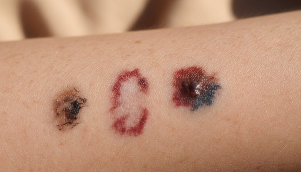
What Does Skin Cancer Look Like – Early Signs, Types & Pictures
Identifying skin cancer in its earliest stages requires understanding how different types manifest on various skin tones and body locations. While many assume all skin cancers present as dark moles, the reality encompasses flesh-colored bumps, persistent scaly patches, and wounds that resist healing.
Medical authorities emphasize that visual characteristics vary significantly between basal cell carcinoma, squamous cell carcinoma, and melanoma—the three primary categories accounting for the majority of diagnoses. What Does Skin Cancer Look Like – Early Signs, Types & Pictures provides additional context for these variations.
Early detection remains crucial, as treatment success rates increase substantially when lesions are identified before spreading occurs.
What Does Skin Cancer Look Like in Early Stages?
| Type | Typical Appearance | Early Warning Signs | Spread Risk |
|---|---|---|---|
| Basal Cell Carcinoma | Fleshy-colored, slightly transparent, shiny bump | New bump that does not disappear over time; occasional ulceration | Low – rarely spreads to other parts |
| Squamous Cell Carcinoma | Red, scaly, crater-like, ulcerated, or bumpy lesions | Wound that never heals; crusty, itchy, may bleed | Moderate – can spread to lymph nodes if untreated |
| Melanoma | Tan, brown, black, or skin-tone/pink (amelanotic) | Asymmetrical changes; rapid color variation | High – can spread to other body areas |
| Actinic Keratosis | Small, dry, scaly, crusty patches with rough texture | Red to dark tan/white/flesh-colored persistent spots | Precancerous – may become SCC |
- Basal cell carcinoma represents the most common type in the UK, presenting as fleshy-colored, slightly transparent bumps that persist over time. Macmillan Cancer Support notes that nearly everyone treated is completely cured.
- Squamous cell carcinoma frequently appears as wounds that never heal, particularly on sun-exposed areas like the scalp and ears.
- Melanoma can develop without pigment (amelanotic), appearing as skin-tone or pink lesions that lack the traditional dark coloration. NHS guidance confirms these variants can appear anywhere on the skin.
- Monthly skin self-examinations significantly improve early detection rates.
- Visual presentation varies considerably based on individual skin tone, lesion size, and specific body location.
- Actinic keratosis serves as a precancerous warning sign, with unpredictable progression to squamous cell carcinoma.
- Persistent itching, crusting, or bleeding from any skin lesion warrants immediate professional evaluation.
| Fact | Details |
|---|---|
| Most common UK skin cancer | Basal cell carcinoma (BCC) |
| Second most common | Squamous cell carcinoma (SCC) – AIM at Skin Cancer |
| BCC growth pattern | Slow-growing, sometimes breaks down and ulcerates |
| SCC typical location | Sun-exposed areas: scalp, ears – Cancer Research UK |
| Melanoma spread risk | Can spread to other body areas |
| Amelanotic melanoma appearance | Skin-tone or pink, lacks pigment – Skin Cancer Foundation |
| Melanoma causes | UV light from sun and sunbeds |
| Actinic keratosis status | Precancerous spots with cells that could become SCC |
| Early detection frequency | Monthly self-examination recommended |
| Treatment success for BCC | Nearly everyone treated is completely cured |
Pictures of Different Types of Skin Cancer
Basal Cell Carcinoma Images
BCC manifests in four distinct presentations. Nodular basal cell skin cancer appears as raised, pearly lesions. Pigmented variants display dark areas, often brown, blue, or grey. Morphoeic basal cell cancer presents as sores that resist healing, accompanied by waxy or scar-like thickened skin. Superficial basal cell skin cancer remains flat or minimally elevated, often resembling red patches.
Squamous Cell Carcinoma Pictures
SCC typically emerges on sun-damaged skin, presenting as red, scaly patches with crater-like centers. These lesions frequently ulcerate and may become bumpy over time. Many patients report persistent crusting and itching, with occasional bleeding upon minimal trauma.
Skin cancers vary considerably due to skin tone, size, type of cancer, and body location. Photographic references serve as guides, not diagnostic tools.
Skin Cancer Appearance on Face, Leg, and Back
Facial Skin Cancer Signs
The face receives disproportionate sun exposure, making it a common site for both basal and squamous cell carcinomas. BCCs on the face often present as shiny, flesh-colored papules with visible blood vessels. SCCs may appear as persistent red patches on the cheeks or nose, frequently crusting but never fully healing.
Leg Lesions to Watch
Lower extremities develop melanoma more frequently than other non-sun cancers, particularly in women. Look for asymmetrical dark patches or amelanotic pink nodules on thighs and calves. Any changing mole or new pigmented spot on the legs requires evaluation.
Back and Trunk Changes
The back presents unique challenges for self-examination, yet hosts significant melanoma risk. Irregularly shaped dark lesions or patches larger than six millimeters warrant attention. Amelanotic melanomas on the back may appear as skin-tone raised bumps rather than dark spots.
Common Symptoms of Skin Cancer
Beyond visual identification, tactile and temporal symptoms provide crucial diagnostic clues. Persistent itching, crusting, or bleeding from any skin lesion indicates potential malignancy. Pain typically manifests only in advanced stages, making visual surveillance essential.
If you see something new, changing, or unusual – even if it looks nothing like photos – do not wait. Early findings and treatment can save your life.
Actinic keratosis appears as small, dry, scaly, or crusty skin patches with rough texture. Clinicians recommend removal because some will become cancer, though you cannot predict which ones will transform.
How Skin Cancer Develops Over Time
- Precancerous Changes: Actinic keratosis develops as small, dry, scaly patches with rough texture, representing cellular damage that could progress to squamous cell carcinoma according to AIM at Skin Cancer.
- Early Basal Cell Carcinoma: Initially appears as fleshy-colored, slightly transparent bumps that do not disappear over time, growing slowly without causing pain.
- Progressing Squamous Cell Carcinoma: Begins as red, scaly patches that may crater or ulcerate; remains relatively slow-growing but can bleed and crust persistently according to Cancer Research UK.
- Advanced Untreated SCC: Can spread to lymph nodes if left untreated for extended periods, though rapid metastasis remains uncommon according to Macmillan Cancer Support.
- Early Melanoma: Develops as new or changing pigmented lesions, sometimes without pigment (amelanotic), capable of spreading to other body areas according to NHS.
Established Facts vs. Uncertain Presentations
| Established Characteristics | Uncertain Presentations |
|---|---|
| BCCs are fleshy-colored, slightly transparent, and/or shiny bumps | Whether specific actinic keratosis lesions will transform into cancer |
| SCCs present as wounds that never heal, often red and scaly | Exact timeline for precancerous progression to invasive cancer |
| Melanoma caused by UV light from sun and sunbeds | Individual genetic predisposition for specific cancer types |
| Risk factors include age, pale skin, and family history | Precise visual differentiation without dermatoscopic examination |
Why Skin Cancer Appearance Varies by Individual
Genetic factors influence how skin cancer manifests across different populations. Melanin levels affect coloration, with darker skin tones sometimes masking early visual signs. Lesion location determines sun exposure patterns, while age impacts cellular repair mechanisms.
The same cancer type presents differently based on body location. Facial skin shows basal cell carcinoma as shiny papules, while back lesions may appear as flat patches. What Does Skin Cancer Look Like – Early Signs Pictures Types illustrates these anatomical variations.
Medical resources including the NHS skin cancer guide provide comprehensive visual references for these variations.
What Medical Authorities Say About Identification
“Skin cancers can look very different and might be: a spot or sore, a lump, a red or dark patch, itchy, crusty or bleeding.”
— Cancer Research UK
“The earlier skin cancer is diagnosed, the easier it is to treat.”
— Cancer Research UK
When to Consult a Dermatologist
Schedule immediate evaluation for any new, changing, or unusual skin changes, particularly those persisting beyond three weeks. Monthly self-examinations facilitate early recognition, while professional dermatoscopy provides definitive diagnosis through biopsy when visual examination proves inconclusive.
Common Questions About Skin Cancer Identification
What does basal cell carcinoma look like?
Basal cell carcinoma typically appears as a fleshy-colored, slightly transparent, shiny bump or lesion that does not disappear over time. Variants include pigmented (dark areas), nodular (raised bumps), and morphoeic (waxy, scar-like) presentations.
What do pictures of squamous cell skin cancer show?
Squamous cell skin cancer pictures typically display red, scaly, crater-like, or ulcerated lesions that may appear bumpy. Many manifest as wounds that never heal, frequently occurring on sun-exposed areas like the scalp or ears.
Is skin cancer on the breast common?
While breast skin can develop melanoma or non-melanoma cancers, it is not among the most common locations compared to sun-exposed areas. Any persistent changes in breast skin, including dimpling or redness, require medical evaluation.
What do skin cancer keratoacanthoma pictures show?
Specific visual references for keratoacanthoma were not identified in current medical databases. Consult dermatological resources or NHS guidelines for authoritative images of this specific lesion type.
How does amelanotic melanoma appear?
Amelanotic melanoma lacks pigment and appears as skin-tone or pink lesions rather than the typical tan, brown, or black coloration. This variant develops anywhere on the skin, including palms, soles, and under nails.
What does skin cancer look like on darker skin tones?
Skin cancers vary considerably by skin tone. On darker skin, melanomas often appear on palms, soles, or under nails, while BCCs may present as darker or pigmented lesions rather than pearly pink bumps.
More related posts
 Camila Mendes Biography: Age, Height, Relationships & Movies
 Secrets Lanzarote Resort & Spa – Adults-Only Reviews and Guide
 Kilimanjaro Peak: Height, Difficulty, Deaths & Beginner Guide
 How to Delete WhatsApp Messages: Complete Guide (2025)
 Does Pepsi Max Have Caffeine? Amount, Comparisons & Sleep Effects
 Secret Santa Gift Ideas: Best Affordable Picks 2024
 Luke Humphries Weight Loss: How 4 Stone Changed His Game
 Satish Shah Dead at 74: Children, Wife’s Alzheimer’s, Legacy
