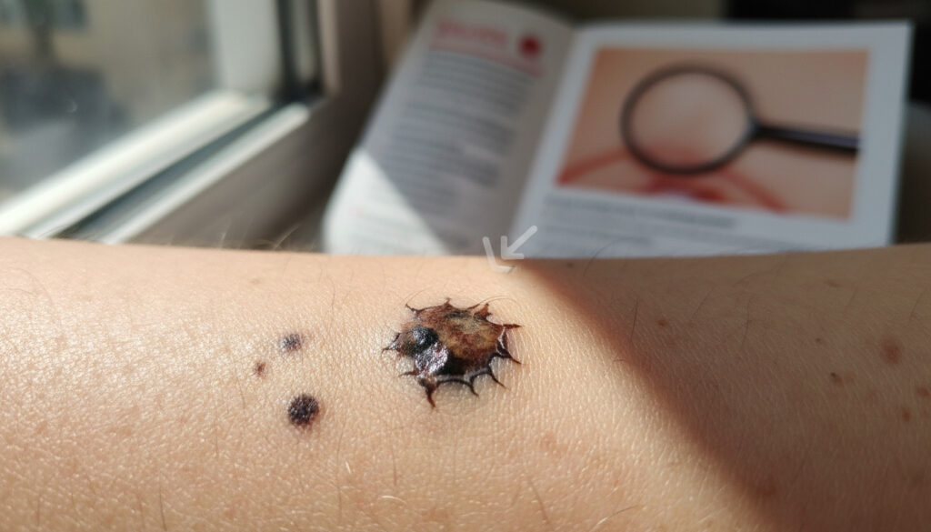
What Does Skin Cancer Look Like – Early Signs and Pictures
Skin cancer rarely announces itself with dramatic symptoms. In early stages, it often appears as subtle changes to the skin—a persistent spot that refuses to heal, a slightly raised bump with an unusual sheen, or a patch of rough texture that lingers for weeks. These initial signs frequently develop on sun-exposed areas, particularly the face, neck, and hands, where ultraviolet radiation has done its cumulative damage over years.
Recognizing these early warnings matters because treatment success depends heavily on timing. While some forms remain locally confined for years, others possess aggressive potential from the outset. Understanding the visual vocabulary of each type—whether basal cell carcinoma’s pearly translucence or melanoma’s irregular borders—enables faster professional evaluation and significantly improves outcomes.
What Does Skin Cancer Look Like in Early Stages?
Early detection relies on recognizing four primary visual categories that distinguish malignant lesions from benign skin changes.
Spots, lumps, and persistent patches that differ in texture or color from surrounding skin, often described as pearly, scaly, or crusty.
Non-healing sores, translucent bumps with visible blood vessels, and scaly areas with raised edges that persist beyond four weeks.
Nose, cheeks, eyelids, and lips where UV exposure concentrates, creating higher risk for basal and squamous cell development.
BCC presents pearly or shiny; SCC appears scaly or crusty; melanoma shows irregular dark pigmentation; keratoacanthoma forms dome-shaped nodules.
Key Insights for Early Recognition
- Basal cell carcinoma accounts for roughly 80 percent of diagnoses and typically grows slowly, rarely spreading to distant organs.
- Melanoma represents the minority of cases but carries the highest mortality risk if detection delays allow metastasis.
- Itching occurs in some basal cell and squamous cell presentations, but absence of itch does not rule out malignancy.
- Squamous cell carcinoma demonstrates higher metastatic potential than basal cell, particularly when appearing on lips or ears.
- Keratoacanthoma variants may spontaneously resolve, yet require biopsy to distinguish from squamous cell carcinoma.
- The ABCDE criteria—Asymmetry, Border irregularity, Color variation, Diameter greater than 6mm, and Evolution—specifically guide melanoma suspicion.
- Early lesions often masquerade as benign pimples or scars, persisting beyond four weeks without improvement.
Visual Characteristics by Type
| Type | Early Appearance | Common Location | Key Symptoms |
|---|---|---|---|
| BCC (Shiny/Pearly) | Translucent bump with waxy or pearly sheen | Face, neck | Bleeding, crusting, occasional itch |
| BCC (Translucent) | Flesh-colored or pink lesion with visible blood vessels | Nose, cheeks | Scabbing, slow growth |
| BCC (Scaly) | Flat patch with raised edges, scar-like appearance | Face, trunk | Persistent scaling |
| BCC (Flat) | Flesh-colored or brown scar-like area | Face, neck | Itch, persistence |
| SCC (Scaly Patch) | Red or pink scaly hyperkeratotic area | Lips, ears, hands | Itch, soreness |
| SCC (Crusty Nodule) | Firm crusted bump or horn-like projection | Sun-exposed skin | Tenderness, bleeding |
| Melanoma | Irregular dark spot with varied pigmentation | Anywhere including face | Evolution in size, shape, or color |
| Keratoacanthoma | Rapidly growing dome-shaped crateriform nodule | Sun-exposed face | Rapid growth over weeks |
Cancer Research UK identifies basal and squamous cell carcinoma as the primary non-melanoma types requiring distinct visual monitoring approaches. Source: Cancer Research UK
What Does Skin Cancer on the Face Look Like?
The face receives more sun exposure than any other body part, making it the most common site for non-melanoma skin cancers. Each subtype tends to favor specific facial zones and presents with location-specific characteristics.
Basal Cell Carcinoma on Facial Skin
On the nose and cheeks, basal cell carcinoma typically manifests as translucent bumps with telangiectasia—visible tiny blood vessels coursing across the surface. These lesions often carry a pearly or waxy quality distinct from surrounding skin. When appearing on eyelids, BCC may present as shiny patches that crust periodically. The ears develop pearly nodules that bleed easily with minimal trauma, a hallmark distinguishing them from benign cysts.
Squamous Cell Carcinoma Presentations
Squamous cell carcinoma favors the lips and ears, where it appears as persistent scaly patches or non-healing sores. Unlike basal cell presentations, SCC on the lips carries significantly higher metastatic potential. On the cheeks, these lesions appear as crusty nodules with a rough, sandpaper-like texture distinguishable from the smooth translucence of BCC. Bowen’s disease—a form of SCC in situ—presents as larger, redder, and scalier patches than actinic keratosis, sometimes causing localized itch or soreness.
Melanoma and Facial Moles
Melanoma on the face appears as irregular dark lesions, either developing suddenly or evolving from existing moles. These spots demonstrate darker pigmentation and more irregular borders than basal cell presentations. While typically pigmented, rare amelanotic variants may appear pink or red, requiring careful border assessment rather than color evaluation alone.
The nose, ears, and lips present particular vulnerability. Squamous cell carcinoma on the lip carries significantly higher metastatic potential than when found on other body areas, requiring immediate evaluation.
The American Society for Mohs Surgery notes that basal cell carcinoma may itch, bleed, crust, heal then recur, or grow slowly—symptoms particularly noticeable on facial skin where patients examine features daily. Source: Mohs Surgery
Pictures of Different Types of Skin Cancer
While visual references guide recognition, remember that variation exists between individuals. The following descriptions correspond to medical imaging databases used for dermatological training and clinical identification.
Basal Cell Carcinoma Gallery
Medical imagery shows basal cell carcinoma as shiny or pearly papules, often described as waxy blue, black, or white lesions depending on depth and pigmentation. Superficial variants appear as flat, flesh-colored or pink scars that lack the translucence of nodular forms. The characteristic rolled border with central depression distinguishes these from benign moles.
Squamous Cell Carcinoma Images
Photographic references display hyperkeratotic red patches that distinguish SCC from actinic keratosis through thickness and persistence. Bowen’s disease presentations appear as larger, redder, and scalier plaques than surrounding sun-damaged skin. Invasive SCC photographs show firm nodules with crusting or ulceration, particularly evident on lip and ear locations.
Melanoma Visual Markers
Clinical images of melanoma reveal dark, irregular spots following ABCDE asymmetry patterns. These lesions show varied pigmentation within single spots, contrasting sharply with the uniform coloration of benign moles. Border irregularity appears jagged or notched rather than smoothly rounded.
Keratoacanthoma Distinctive Features
Visual databases identify keratoacanthoma as dome-shaped nodules with central keratin-filled craters, resembling small volcanic structures. The rapid growth phase—often photographed in sequence over weeks—distinguishes them from the slower development typical of basal cell carcinoma.
What Does Skin Cancer Look Like – Early Signs, Types and Pictures provides additional visual references for comparative self-assessment.
The American Cancer Society describes squamous cell carcinoma as originating from upper epidermis squamous cells, presenting visually as red or pink scaly patches or crusty bumps distinct from basal cell origins. Source: American Cancer Society
Skin Cancer Symptoms to Watch For
Beyond visual appearance, specific physical sensations and behavioral patterns indicate malignant processes requiring investigation.
Itching and Sensory Changes
Itch presents in developing basal cell carcinoma and Bowen disease presentations of squamous cell carcinoma, though not universally. Tenderness or soreness occurs more frequently in SCC than BCC, particularly as lesions thicken or ulcerate. Any persistent unexplained sensation in a specific skin area warrants examination.
Bleeding and Surface Changes
Spontaneous bleeding from minimal trauma distinguishes cancerous lesions from benign growths. Crusting that recurs after falling off, or non-healing sores persisting beyond four weeks, represents classic warning signs. These symptoms appear across BCC and SCC types, particularly on facial skin exposed to daily washing and shaving trauma.
Evolution and Growth Patterns
Rapid growth over weeks suggests keratoacanthoma or aggressive squamous cell carcinoma, contrasting with the slow, steady expansion typical of basal cell carcinoma. Any lesion doubling in size within three months requires immediate dermatological assessment.
Visual assessment alone cannot confirm malignancy. Biopsy remains the definitive diagnostic tool. Suspicious lesions require dermatological evaluation regardless of symptom presence or absence.
Any skin change that persists longer than one month without healing warrants professional examination, particularly on sun-exposed facial areas.
MD Anderson Cancer Center emphasizes that early symptoms overlap across types, with itching, pain, and bleeding occurring in growing BCC and SCC patches, while melanoma demonstrates evolution in appearance. Source: MD Anderson Cancer Center
How Does Skin Cancer Progress Over Time?
Understanding the temporal behavior of each cancer type clarifies why some require urgent intervention while others allow scheduled monitoring.
- Initial Development (Months 1–6): UV-damaged cells begin uncontrolled division. BCC forms pearly bumps; SCC creates scaly patches; melanoma develops from atypical moles or new pigmented lesions.
- Local Growth (Months 6–24): BCC slowly expands locally, rarely breaking deep into underlying tissue. SCC penetrates deeper epidermal layers, risking lymph node involvement if untreated. Melanoma enters vertical growth phase with potential for early metastasis.
- Regional Spread (Year 2–5): Untreated SCC spreads to regional lymph nodes in 2–5 percent of cases. Melanoma metastasizes to distant organs, dramatically reducing survival rates once beyond local skin layers.
- Advanced Disease: BCC causes disfiguring local destruction if neglected over years. SCC may cause significant morbidity through metastasis. Melanoma becomes systemic, affecting organs and survival probability.
Dermatology Seattle notes that melanoma proves most deadly due to metastasis capability, while basal cell carcinoma rarely spreads but remains locally destructive. Source: Dermatology Seattle
What Medical Science Confirms vs. What Remains Uncertain
Distinguishing established diagnostic criteria from areas requiring further research helps set realistic expectations for self-monitoring and clinical evaluation.
Established Facts
- BCC presents as pearly, translucent, or scar-like lesions in the majority of cases
- SCC originates in upper epidermis with scaly, crusty appearance
- Melanoma follows ABCDE detection criteria with high reliability
- UV exposure directly causes DNA damage leading to these cancers
- Early surgical treatment cures the majority of non-melanoma cases
Uncertain or Variable
- Exact progression timeline without treatment varies individually by immune response
- Why some itchy lesions turn cancerous while others remain benign
- Specific visual differentiation between keratoacanthoma and SCC without biopsy
- Whether all keratoacanthoma cases would eventually metastasize if untreated
- Precise visual appearance variations across all skin tones (underrepresented in early studies)
Calderm Institute research highlights that squamous cell carcinoma grows faster and can metastasize, contrasting with basal cell carcinoma’s slower, locally confined behavior. Source: Calderm Institute
Why Skin Cancers Look Different by Type
The cellular origin determines the visual presentation. Basal cell carcinomas emerge from the epidermis’s deepest layer, creating translucent, slow-growing bumps that maintain structural organization despite malignancy. Squamous cell carcinomas arise from upper-layer keratinocytes, producing the rough, scaly texture associated with surface skin disruption.
Melanomas originate from pigment-producing melanocytes, explaining their characteristic dark, irregular appearance as these cells continue producing melanin erratically. This biological foundation explains why a pearly bump differs fundamentally from a dark spreading spot, despite both representing malignant processes requiring intervention.
Bare Derm research confirms that melanoma’s aggression and metastasis risk distinguish it from carcinomas, explaining why visual identification carries different urgency levels. Source: Bare Derm
What Medical Authorities Say About Identification
Skin cancers in early stages on the face often appear as small, persistent lesions in sun-exposed areas, with basal cell carcinoma showing as shiny or pearly bumps, squamous cell carcinoma as scaly red patches, and melanoma as irregular dark spots.
MD Anderson Cancer Center
Basal cell carcinoma starts in the basal cells of the epidermis’ lower layer… may itch, bleed, crust, heal then recur, or grow slowly.
American Society for Mohs Surgery
Melanoma is the rarest but deadliest, aggressive with high metastasis risk if advanced.
Dermatology Seattle
The American Academy of Dermatology maintains that recognizing these distinct presentations across types remains essential for early detection and treatment success. Source: American Academy of Dermatology
When to Consult a Dermatologist
Schedule an appointment when you notice any new growth that persists beyond four weeks, changes in existing moles following the ABCDE criteria, or non-healing sores on sun-exposed skin. While basal cell and squamous cell carcinomas rarely prove fatal when caught early, melanoma demands immediate attention. Regular skin examinations, combined with professional assessment of suspicious changes, provide the strongest protection against advanced disease. What Does Skin Cancer Look Like – Key Signs to Spot Early offers additional guidance on preparing for your appointment.
Frequently Asked Questions
Are NHS skin cancer images different from other medical photos?
NHS databases contain the same diagnostic imagery used internationally, though UK-specific resources may emphasize presentations more common in the British population. Consult NHS.uk or Cancer Research UK galleries for reference.
Does skin cancer always itch?
No. While itching occurs in some basal cell carcinomas and Bowen disease presentations, many malignant lesions remain asymptomatic. Absence of itching does not indicate absence of cancer.
Can skin cancer look like a pimple?
Yes. Early basal cell carcinoma may resemble a persistent pimple or shiny bump that never resolves. Unlike acne, these lesions remain for months and may bleed with minimal trauma.
How quickly does keratoacanthoma grow?
Keratoacanthoma typically grows rapidly over weeks, forming dome-shaped nodules. This contrasts with basal cell carcinoma’s slower progression. Biopsy is essential to distinguish it from squamous cell carcinoma.
Is every scaly patch on the face skin cancer?
No. Actinic keratosis presents as scaly patches but remains pre-cancerous. However, Bowen’s disease appears similarly and requires treatment. Only biopsy confirms the diagnosis.
Why does melanoma look more irregular than other skin cancers?
Melanoma arises from melanocytes that produce pigment irregularly as they become malignant, creating varied colors and borders. This irregularity contrasts with the uniform pearly appearance of basal cell carcinoma.
More related posts
 Area of a Sector: Formula, Calculator & Step-by-Step Guide
 UK Weather Tornado Warning – No Active Alerts Today
 Flats to Rent Near Me in Ireland: Daft.ie Listings
 Me and Em UK – Stores, Online and Returns Guide
 Club Brugge vs Rangers F.C. Lineups – Confirmed and Predicted XIs
 Toyota Yaris for Sale in Ireland: Used & New Deals
 Paul Gascoigne 2025: Health, Sobriety, Net Worth Update
 UK Driving Theory Test – Book, Practice & Pass Guide 2025
