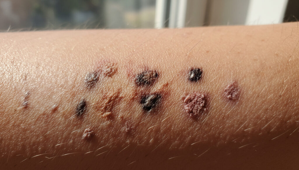
What Does Skin Cancer Look Like – Early Signs Pictures Types
Skin cancer in its early stages typically manifests as new, changing, or unusual spots, sores, lumps, or patches on the skin. These changes frequently appear on sun-exposed areas such as the face, legs, and back, though they can develop anywhere on the body. Recognizing these visual warning signs promptly significantly improves treatment outcomes.
Medical image galleries from organizations including the American Cancer Society and the Skin Cancer Foundation document diverse presentations across different skin tones and body locations. While common types including basal cell carcinoma, squamous cell carcinoma, and melanoma each display distinct visual characteristics, early detection remains the critical factor regardless of specific appearance.
What Does Skin Cancer Look Like – Early Signs, Types & Pictures provides additional visual reference materials, while this guide examines photographic evidence from leading dermatological sources to illustrate how skin cancer presents in early stages, across different body parts, and among various subtypes.
What Does Skin Cancer Look Like in Early Stages?
Early-stage skin cancers typically begin as small, evolving lesions that may be mistaken for benign conditions. Cancer Research UK photographic archives indicate that initial presentations vary significantly by cancer type and individual skin characteristics.
| Common Appearances | Color Indicators | Texture Changes | Detection Framework |
|---|---|---|---|
| Spots, lumps, patches, or non-healing sores | Red, brown, purple, black, or pearly translucent | Crusty surfaces, itching, spontaneous bleeding | ABCDE rule assessment for asymmetry and evolution |
- Early lesions often start smaller than 6mm but evolve gradually over weeks or months
- Not all skin changes indicate malignancy, but persistence beyond three weeks warrants evaluation
- Visual patterns differ significantly: BCC appears pearly, SCC presents as scaly patches, melanoma shows multi-colored asymmetry
- Sun-exposed areas demonstrate higher frequency of occurrence but cancers may develop on any skin surface
- Skin tone affects color perception; on darker skin, BCC may appear flesh-colored or translucent rather than pink
- The ABCDE rule provides a systematic framework for distinguishing suspicious lesions from benign moles
- Monthly self-examination improves early identification rates substantially
| Type | Early Appearance | Common Location |
|---|---|---|
| Basal Cell Carcinoma (BCC) | Pearly or waxy bump, shiny nodule, pinkish patch | Face, ears, neck, scalp |
| Squamous Cell Carcinoma (SCC) | Scaly, rough, red patches or non-healing sores | Face, ears, lips, hands, legs |
| Melanoma | Changing mole >6mm with uneven color, irregular borders | Back, legs, neck, anywhere including soles |
| Keratoacanthoma | Dome-shaped crater with central keratin plug | Sun-exposed face, arms |
| Actinic Keratosis (Precancerous) | Dry, scaly, rough spots in red, tan, or white | Sun-exposed areas |
| Nodular BCC | Raised shiny bump with visible blood vessels | Face, head |
| Morphoeic (Sclerosing) BCC | Scar-like or slowly enlarging sore | Face, trunk |
| Superficial BCC | Flat, scaly, pink or red patch | Back, legs |
What Does Skin Cancer Look Like on the Face, Leg, or Back?
Anatomical location influences both the typical presentation and visibility of skin cancer. Mohs College photographic resources demonstrate that facial lesions often differ in morphology from those appearing on the lower extremities or back.
Facial Presentations
The face, particularly the nose and ears, commonly develops basal cell carcinomas presenting as pearly nodules or pinkish bumps. Squamous cell carcinoma on the face typically manifests as scaly patches or firm pink bumps that may crust or bleed. These lesions often appear on the lips, ears, or scalp in individuals with significant sun exposure history.
Pay particular attention to the eyelids, ears, and lips—areas where skin cancers may develop distinct characteristics such as loss of normal skin lines, persistent redness, or small ulcers that fail to heal within four weeks.
Lower Extremity Indicators
Legs frequently harbor melanomas, particularly in women, where lesions may appear as irregularly colored spots on the calves or thighs. Basal cell carcinoma on the legs often presents as flat, scaly patches rather than the pearly bumps seen on the face. Squamous cell carcinoma may appear as rough, scaly areas or non-healing sores on sun-exposed thighs and lower legs.
Back and Torso Signs
The back represents a common site for melanoma development, where lesions often go unnoticed due to limited visibility. These may appear as changing moles or new pigmented spots >6mm with asymmetrical borders and color variation. Basal cell carcinomas on the back typically present as flat, scaly patches or pinkish lesions, while squamous cell carcinomas manifest as rough, scaly plaques or red patches.
Pictures of Different Types of Skin Cancer
Mayo Clinic dermatological archives categorize skin cancers by cellular origin and growth patterns, each producing distinct visual signatures that aid in preliminary identification.
Basal Cell Carcinoma Visual Characteristics
As the most common form, BCC presents multiple subtypes. Nodular BCC appears as a raised, translucent, pearly bump with visible telangiectasia (tiny blood vessels). Morphoeic or sclerosing BCC resembles a scar or white, waxy, sun-damaged area that slowly enlarges. Pigmented BCC displays brown, blue, or black tones, potentially mimicking melanoma. Superficial BCC spreads horizontally as a flat, scaly, pink or red patch, often on the trunk or limbs.
Squamous Cell Carcinoma Appearance
SCC in situ (Bowen’s disease) presents as persistent, scaly, reddish patches. Invasive SCC may appear as firm, raised lumps with a central crust or ulceration, sometimes forming horn-like growths. American Academy of Dermatology images show these lesions frequently occurring on the rim of the ear, face, neck, hands, or arms.
Melanoma Recognition Patterns
Melanoma demonstrates the most variable appearance, earning its reputation as the most visually deceptive skin cancer. The “ugly duckling” sign—where one lesion differs significantly from a person’s other moles—serves as a critical warning. Amelanotic melanoma appears pink or red rather than pigmented, while nodular melanoma grows vertically as a firm, dome-shaped bump. University of Texas Southwestern Medical Center documentation emphasizes that melanoma may occur on palms, soles, or under nails, appearing as streaked discolorations.
Keratoacanthoma Distinct Features
This rapidly growing tumor mimics SCC, appearing as a dome-shaped nodule with a central keratin plug that creates a volcano-like crater. While some spontaneously resolve, they require professional evaluation to exclude squamous cell carcinoma. These typically emerge on sun-exposed facial skin or forearms.
Skin Cancer Symptoms and NHS Images
The UK’s National Health Service and WebMD clinical resources emphasize that visual changes often accompany physical symptoms indicating potential malignancy.
The ABCDE Rule for Melanoma Detection
AIM at Skin Cancer and NHS resources promote the ABCDE mnemonic for systematic assessment:
- Asymmetry: One half of the lesion does not match the other
- Border: Irregular, notched, or blurred edges rather than smooth, even borders
- Color: Variable shades including brown, black, red, white, or blue within a single lesion
- Diameter: Greater than 6mm (pencil eraser size), though smaller melanomas occur
- Evolving: Changes in size, shape, color, elevation, or new symptoms such as bleeding, itching, or crusting
When Symptoms Indicate Urgent Evaluation
Persistent itching, tenderness, bleeding without trauma, or non-healing sores lasting longer than four weeks require dermatological assessment. Physical sensations often distinguish malignant lesions from benign growths like seborrheic keratoses, which typically remain stable and asymptomatic.
Image galleries provide reference examples only; definitive diagnosis requires histopathological examination. Some malignant lesions resemble benign conditions, and vice versa. Professional evaluation remains essential for any suspicious or changing skin lesion.
NHS and Cancer Research UK photographic databases undergo clinical validation by dermatologists, providing trustworthy visual references for non-melanoma and melanoma skin cancers across diverse skin tones.
How Does Skin Cancer Progress Over Time?
Understanding the temporal evolution of skin lesions helps distinguish transient benign conditions from persistent malignancies requiring intervention.
- Initial Presentation: Small, often overlooked spot or patch with subtle color or texture changes
- Surface Evolution: Gradual development of distinct morphological features—pearly translucency, scaliness, or multi-coloration—over weeks to months
- Structural Changes: Emergence of raised borders, central ulceration, or nodularity indicating vertical or invasive growth
- Symptom Development: Onset of spontaneous bleeding, persistent crusting, itching, or tenderness not triggered by external irritation
- Advanced Characteristics: Significant size increase (>6mm), irregular border definition, or satellite lesions surrounding primary growth
What Can Visual Assessment Confirm Versus What Requires Clinical Verification?
Photographic references provide valuable screening tools, yet certain diagnostic certainties remain exclusively within clinical scope.
| Established Visual Indicators | Information Requiring Clinical Confirmation |
|---|---|
| Lesion asymmetry and border irregularity observable through comparison | Definitive malignancy versus benign mimic without histopathological biopsy |
| Color variation and diameter exceeding 6mm measurable by physical inspection | Specific cancer subtype classification and histological grading |
| Evolving characteristics documented through serial photography | Full staging, depth of invasion, and metastatic potential assessment |
| Sun-damaged skin areas with multiple actinic keratoses | Which specific lesions require treatment versus monitoring |
What Factors Influence How Skin Cancer Appears?
Individual characteristics significantly modify the visual presentation of skin malignancies. Skin tone profoundly affects lesion coloration; basal cell carcinomas on darker skin may present as translucent, pearly, or flesh-colored rather than the typical pink or red seen on lighter skin. This variation can delay recognition without careful examination.
Sun exposure history, age, and genetic predisposition also determine lesion location and morphology. Chronic UV damage typically produces lesions on the face, neck, and hands, while intermittent intense exposure correlates with trunk and leg melanomas. Immunosuppressed individuals may develop more aggressive-appearing lesions with atypical coloration.
Previous skin cancer history increases the likelihood of new primary lesions, requiring heightened surveillance for any new or changing spots regardless of initial benign appearance.
What Do Leading Medical Authorities Say About Visual Detection?
Reputable medical organizations emphasize the importance of structured visual assessment while cautioning against reliance solely on photographic comparisons.
“Examine skin monthly; see a doctor or dermatologist for anything new, changing, or unusual, even if unlike gallery images—early detection cures most skin cancers.”
— Clinical guidelines from comprehensive dermatological resources
“Mohs surgery provides effective treatment for basal and squamous cell carcinomas, while precancerous conditions like actinic keratosis warrant removal before progression.”
— Dermatologic surgery societies
What Are the Essential Visual Indicators to Remember?
Skin cancers present diverse visual signatures—pearly bumps, scaly patches, evolving moles, or crater-like nodules—requiring vigilant monitoring rather than categorical memorization. The fundamental principle remains consistency: any lesion demonstrating asymmetry, border irregularity, color variation, diameter increase, or evolution demands professional evaluation regardless of whether it matches typical photographic examples. What Does Skin Cancer Look Like – Key Signs to Spot Early offers additional guidance on recognizing these critical changes.
Frequently Asked Questions
What does basal cell carcinoma look like specifically on the face?
Facial BCC typically appears as a pearly or waxy bump, often with visible small blood vessels, or as a flat, flesh-colored or pinkish patch resembling scar tissue. On the nose and ears, it may present as a shiny, translucent nodule that occasionally bleeds or crusts.
Is every crusty spot on the leg potentially skin cancer?
Not all crusty leg lesions indicate malignancy; eczema, psoriasis, or insect bites may produce similar appearances. However, persistent scaly patches lasting longer than three weeks, especially on sun-exposed thighs or calves, require dermatological evaluation to exclude squamous cell carcinoma or actinic keratosis.
How can I distinguish melanoma from a normal mole using pictures?
Melanoma typically displays the ABCDE characteristics: asymmetry (two halves differ), irregular borders, multiple or uneven colors, diameter larger than 6mm, and evolution over time. The “ugly duckling” test—one lesion looking different from your other moles—also indicates potential concern.
What does keratoacanthoma look like compared to squamous cell carcinoma?
Keratoacanthoma grows rapidly as a dome-shaped, volcano-like nodule with a central keratin plug, often on sun-exposed facial skin or arms. While visually similar to SCC and sometimes classified as a variant, it may spontaneously resolve, though surgical removal remains necessary for definitive diagnosis and treatment.
Are NHS skin cancer images reliable for self-diagnosis?
NHS and Cancer Research UK images provide clinically validated, trustworthy visual references across diverse skin tones and body locations. However, they serve as educational tools rather than diagnostic instruments; any suspicious lesion requires professional medical assessment regardless of photographic similarity.
Why does skin cancer look different on darker skin tones?
Melanin concentration affects lesion coloration; BCC may appear translucent, gray, or flesh-colored rather than pearly pink on darker skin. SCC might present as raised, scaly, or hyperpigmented lesions. This variation necessitates careful examination beyond typical textbook examples to avoid delayed diagnosis.
What visual signs indicate a sore is cancerous rather than healing normally?
Non-healing sores persisting beyond four weeks, particularly those that bleed spontaneously, develop raised or rolled borders, or show central crater formation, suggest malignancy. Cancerous lesions typically lack the uniform contraction and granulation tissue seen in normal wound healing.
More related posts
 Mlp Fluttershy: Complete Guide to the Shy Pegasus
 Jodie Whittaker: Children, Husband, New Look & Frauds
 The Range Discount Code – Verified UK Deals and Student Savings
 Where Is the Liver Located? Anatomy, Pain & Functions
 Companies House Identity Verification Launch – Dates Process and Requirements
 Pain Lower Left Abdomen: Causes in Males & Females
 Tana Mongeau: Fame, Net Worth, OnlyFans, & Controversies
 Lash Extensions Near Me: Prices, Salons & Care in Ireland
